
News
April 2015: Alex Rauscher is Canada Research Chair in Developmental Neuroimaging
May 2014: Vanessa Wiggermann recieves 2nd place ISMRM White Matter Study Group Award 2014
March 2014: Vanessa Wiggermann recieves doctoral studentship from the MS Society of Canada (renewed in 2015)
March 2014: Alexander Wright receives a Vanier Scholarship
March 2014: Evan Chen receives Frederick Banting and Charles Best Canada Graduate Scholarship 2014/15
April 2013: Evan Chen wins the ISMRM White Matter Study Group Presentation Award.
April 2013: Alexander Rauscher received the London Drugs Award for Radiology Research Excellence
April 2013: Enedino Hernandez, Vanessa Wiggermann and Evan Chen received ISMRM Stipend Awards to attend the annual meeting in Salt Lake City.
June 2012: Alex Rauscher receives CIHR New Investigator Award
June 2011: Glen Foster receives Michael Smith Postdoctal Fellowship
Funding
Canada Research Chair
CIHR New Investigator Award (2012-2017)
NSERC, Parkinson Society Canada, UBC Spring Startup Fund (PI);
CIHR, MS Society, National Parkinson Foundation (co-investigator)
Current Projects
Sports Concussion
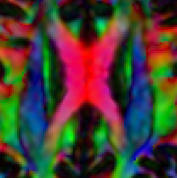
We are conducting a study on sport concussion using neuroimaging and neuropsychological testing. We are following two ice hockey teams over the 2011/12 season. All hockey players have received baseline assessment including MRI and neurospychological testing. Players who have a concussion receive serial tests over two months after the concussion. We are using advanced MRI techniques, such as multi echo SWI for the detection of brain haemorrhages, and diffusion tensor imagiging to assess changes in the brain due to concussion.
MR Frequency Shifts
In this study funded by NSERC we investigate the MR signal of anisotropic tissues.
Multiple Sclerosis
We use MR frequency imaging for the investigation of tissue changes due to multiple sclerosis. In a serial study, we found that new MS lesions formation leads to an increase in MR frequency.
Spinal Cord
We use MR frequency imaging for the investigation of spinal cord injury.
General Research Interests
Phase Information of the MR Signal
Magnetic resonance imaging (MRI) is
a tremendously successful tool for biomedical research and diagnostic
imaging. MRI research resides at the interfaces of life sciences and
natural sciences and their respective subdisciplines.
Most of the
information gathered with MRI is due to subtle variations in signal
magnitude which can be translated into images with meaningful
information about the nuclei's biophysical environment. The MRI
signal, however, has another, promising, yet underexploited property:
phase. The phase is best explained by using the analogy of a
lighthouse: The MRI signal's magnitude corresponds to the brightness
of the lighthouse beam and the MRI signal's phase corresponds to the
beam's direction. Just as the lighthouse beam always has a certain
direction, the MR signal always has a certain phase. One challenge
with phase is that we do not yet fully understand the underlying
biophysical processes leading to phase information. The conventional
contrasts have been investigated for decades and are, although still
not fully understood, used extensively in MRI. Phase may allow us to
glean further information on tissue composition
and structure.
Applications
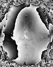
We use the phase's sensitivity to iron for the imaging of iron rich brain strcutures. This allows us the visualization of areas that are difficult to image with conventional MRI. One example is the subthalamic nucleus subthalamic nucleus, which is a target of deep brain stimulation in Parkinson Disease. It was shown that iron is elevated in deep gray matter structures of the brains of people with multiple sclerosis or Parkinson disease. We use our multi echo SWI technique to map these structures.
MR Data Processing
We have developed a region growing algorithm that uses a combination of data quality criteria extracted from the complete complex information. In MRI this is important is we want to preserve all spatial frequencies. Temperature mapping or maps of the magnetic field are examples for such applications. The classic approach in phase imaging, homodyne, filtering does not preserve all spatial frequencies. Usually homodyne filtering is good enough, but in some situations phase unwrapping is superior. One example is venography in areas with strong field inhomogeneities. Another example is mapping of the R2* decay, for example with multi echo SWI, where the correction for unwanted signal decay due to background field inhomogeneities requires field maps.
Software
Phase Unwrapping with PhUN
Our phase unwrapping algorithm is written in C and is available for
Linux, Mac and Windows. It can be called from Matlab, IDL or the
command line. Typical unwrapping times are 0.1 s for a 512 x 512
image.
We are happy to
share this phase unwrapping method. Please
contact Stephan
Witoszynskyj or Alexander Rauscher, if you are
interested.
Lesion Tool
We (Christian Kames, Stephanie Schoerner, Enedino Hernanez-Torres) developed a lesion marking tool that has been useful in a range of studies. If you are interested, please contact us.